The heart is one of the most important organs in the body as it provides oxygen and nutrients to all body tissues.
Along with the brain, kidneys, lungs, and liver, the heart is considered one of the body’s vital organs due to its key role in sustaining life. When a problem occurs with the heart then the rest of the body invariably suffers. Tissues may receive insufficient oxygen or nutrients while metabolic waste and other toxins can build up in organ systems or other locations in the body due to improper draining through the venous (veins) and lymphatic systems. Let’s start with the basic anatomy of the heart and circulatory system and then move onto heart disease causes, diagnosis, treatment, and prevention.
What is Heart Disease?
Heart disease occurs when the heart improperly forms or is damaged in some capacity that affects its function. A problem affecting the heart, circulatory or lymphatic systems can negatively impact whole-body health.
The Underlying Causes of Heart Disease Are Various and Include:
Congenital - Congenital heart disease occurs when the heart improperly forms in utero leading to a pet being born with an abnormally functioning heart. Common congenital heart diseases include Patent Ductus Arteriosus (PDA), Pulmonic Stenosis (PS), and Subaortic Stenosis (SAS). There can be a genetic component to congenital heart disease permitting the passage from an older to younger generation. Certain breeds predisposed to congenital heart disease include the Boxer, Cavalier King Charles Spaniel, Dachshund, Doberman Pinscher, Golden Retriever, Poodle (miniature and toy), Schnauzer (miniature) and mixes of these breeds. Degenerative - As the body ages, tissues will invariably lose their functional capacity to perform at an optimal level. Degenerative Mitral Valve Disease (DMVD) is one type of heart disease that can occur simply as a result of a pet living years of life and can be exacerbated by the presence of other heart ailments or inciting causes (infectious, toxic, etc.). Infectious - Infectious organisms like bacteria, viruses, and others can get into the body and negatively impact the heart. The mouth is one location where bacteria enters the circulatory system through inflamed gum tissue (gingivitis), fractured teeth, or penetrating injury. Blood borne-bacteria may damage normal heart tissue, such as the valves, and cause or worsen heart disease. Toxic - The heart is susceptible to certain man-made and environmental toxins, including plants like Foxgloves, Common and Yellow Oleander and Mistletoe, and chemotherapeutic medications like Adriamycin (Doxirubicin). The degree of toxicity such plants and medications cause to the heart depends on the type, volume, and duration of which such toxins are ingested or injected. Ingestion of stimulants like caffeine, theobromine, cocaine, methamphetamine, pseudoephedrine, and others cause elevated heart rate and blood pressure and can have a toxic effect on the heart and circulatory system. Overproduction of the body’s own thyroid hormone (hyperthyroidism) or excessive supplementation when treating hypothyroidism can cause an abnormal thickening of heart muscle called thyrotoxic cardiomyopathy. Traumatic - When the heart incurs blunt-force trauma, such as that occurring when a pet is hit by a car, takes a fall, or receives a kick or other forcible blow from another creature then bruising can occur that negatively impacts heart function.
Clinical Signs of Heart Disease Can be Mild to Severe and Include:
Lethargy - Heart disease causes all organs to suffer from reduced oxygenation and nutrient delivery and decreased removal of toxins and metabolic wastes and can cause a pet to appear lethargic (tired), be less active, and show exercise intolerance. The degree to which a pet shows signs of lethargy depends on the severity and duration of heart disease. Coughing - When heart disease progresses from mild to severe, there is often heart enlargement (cardiomegaly) which can press on the trachea (windpipe) inside of the chest and cause coughing. Additionally, as heart disease enters the later stages pulmonary edema (fluid in the lungs) can occur which causes a moist-sounding cough and respiratory sounds. Increased respiratory rate and effort - Over time, pets having heart disease will show increased respiratory rate and effort. Increased respiratory rate can be seen and heard by watching and listening to your pet. Normal respiratory rate is quite variable and ranges from 10-30 breaths per minute in dogs and 20-30 breaths per minute in cats. Tachypnea is the term for elevated respiratory rate, bradypnea indicates decreased respiratory rate, and dyspnea means labored or difficulty breathing (gasping, sneezing, spasming, wheezing, etc.). Your pet’s respiratory rate can be calculated by watching the movement of the ribs (expand/contract) and abdomen (in/out). Alternatively, a finger or mirror can be placed in front of nostrils to feel each breath on the fingertip or count the number of times condensation is seen on the mirror with each exhale. Make sure to check both the R and L nostril for air flow. I generally recommend counting breaths for 30 seconds and multiplying by two to calculate the number of breaths per minute. Respiratory effort can be determined by watching and listening to your pet’s breaths. Normal respiratory effort occurs in a consistent and smooth manner with minimal movement of the abdomen. Significant abdominal movements, extension of the neck, movement of the elbows away from the body (abduction), appearance of gasping for breaths, and harsh sounds or wheezing all indicate increased respiratory effort. Abnormal mucus membranes and prolonged capillary refill time - When heart disease causes insufficient oxygen to enter the body, one of the primary places owners can see the difference is in a pet’s mucus membrane (gums, gingiva). Normal mucus membranes appear pink and moist, while abnormal mucus membranes look to be pale, white, blue, grey, purple, brick red, or other unusual color and feel dry or tacky. Capillary referral time (CRT) is the amount of time needed for the color to return to the mucus membranes upon being pressed to push out the blood to induce pallor (pale color). Normal CRT is 1-2 seconds. CRT is also an indicator of hydration, as dehydration causes blood to thicken and flow more slowly to all body parts and will delay CRT. Decreased appetite and weight loss or gain - When heart disease advances, appetite often decreases. As a result, weight loss generally ensues. Yet, some advanced heart diseases can cause fluid to be retained in the abdominal cavity (ascites) and other tissues and lead to weight gain. Collapsing episodes - Pets become weak from progressive heart disease and are less able to stand and participate in their usual activities like walking, running and playing and are prone to collapsing episodes which can appear consistent with fainting (syncope). Edema - With heart disease comes abnormal movement of fluid around the body through the circulatory and lymphatic systems. As a result, fluid can accumulate in the skin of limbs, underside of the chest and abdomen, and other body parts and cause a sponge-like consistency called edema.
How is Heart Disease Diagnosed?
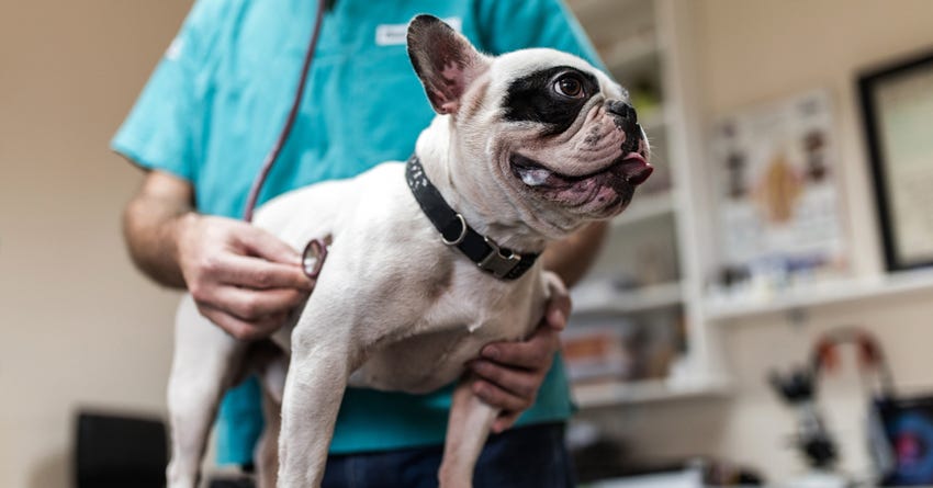
Heart disease is diagnosed a variety of ways and the starting off point is an examination by a veterinarian. It is always best that the diagnosis of heart disease is made before clinical signs occur, so the pet minimally suffers from illness and the likelihood of the condition being managed is greater. Physical examination - When a veterinarian listens to your pet’s heart with a stethoscope (auscultation), then abnormal heart sounds like a murmur or arrhythmia may be heard. A murmur occurs when one or multiple heart values improperly function and don’t do their job in preventing blood from flowing in an abnormal direction between the atria and ventricles. Murmurs are given a grade of 1 through 6 with the severity increasing with higher grade. An arrhythmia is heard when the rhythm with which the heart contracts doesn’t follow a consistent pattern. Bradycardia is an arrhythmia where the heart beats too slow and tachycardia occurs when the heart beats too fast. Additional types of arrhythmias include premature ventricular contractions(PVCs), atrial fibrillation (“AFib”), heart block, and others. Your veterinarian will also feel your pet’s pulses to see if the pace of blood pumping in the arteries matches the sound of the heart beating. The number of beats per minute (BPM) can be assessed by feeling your pet’s arteries. Normal heart rate ranges depending on a pet’s size, hydration, overall health, medication regimen, and lifestyle factors and is 70-160 beats per minute in dogs and 160-240 beats per minute in cats. Smaller creatures typically have higher heart rates than larger animals. Owners can determine the pet’s heart rate by feeling the pulse of blood through the body’s arteries, which is most-easily found at the femoral artery on the inner and upper aspect of the hind limb (femur). A person’s index and middle fingers are used to gently press down on femoral artery until pulse is felt. I recommend counting the number of pulses over 30 seconds and multiplying the number by two to determine the rate over one minute. Counting heart rate through pulse palpation is preferred to putting hand over heart, as it’s too simple to mistake the “lub dub” sounds of the heart’s contraction and relaxation as two heart beats. Evaluation of your pet’s respiratory rate and effort and auscultation of the trachea and lungs are other key components of the physical exam. Electrocardiogram - The heart’s electrical activity is determined by a test called an electrocardiogram (ECG). Electrical impulses that course through the muscular walls of the heart elicit the stimulus to contract and relax so the heart can empty and fill with blood. ECG creates a visual representation of the electrical rhythm of each heart beat and is used to establish if an arrhythmia is present. ECG is non-invasive and simply requires mild restraint on an awake animal, so a series of wired clips can be connected the body’s limbs. Echocardiogram - To determine exactly what parts of the heart are affected and the degree to which malfunctioning occurs, ultrasound imaging is essential. Echocardiogram is an ultrasound image of the heart and certain parts of the circulatory system which creates a real-time series of moving images about heart valve function, blood flow, heart rate, contractility, and more. Like ECG, echocardiogram is non-invasive and is performed on an awake animal using mild restraint. The lower sides of the right and left chest are often clipped free of hair to permit the ultrasound probe to have improved contact with the skin surface to yield better visualization of the heart and its associated structures. Blood pressure - Heart disease can cause an increase or decrease in blood pressure pending the nature of the ailment. Hypertension means increased blood pressure and hypotension indicates reduced blood pressure. Blood pressure determination involves a pressure cuff being placed around a limb which is inflated to restrict blood flow and a stethoscope or ultrasonic device called a Doppler reads the pulse of the returning flow as the cuff is deflated. Attaining a blood pressure requires minimal restraint on an awake animal. Pets that are excited, stressed, or anxious and those having recently been physically active may show elevated blood pressure that is not truly representative of hypertension. Radiographs (x-rays) - Thoracic radiographs reveal static images of the chest cavity‘s contents, including the heart, lungs, blood vessels leading to and from the heart and lungs, and more. Abdominal radiographs may also be needed to evaluate the liver and other organs and to look for ascites and other abnormalities potentially associated with heart disease. Radiographs are minimally invasive and require short-term mild restraint for proper positioning to attain the best-possible images. Blood testing - Creatinine kinase (CK, a muscle enzyme), IDEXX Laboratories Pro BNP (a substance “released by cardiac myocytes in response to stretch and stress”), electrolytes, calcium, blood proteins (albumin, etc.), kidney and liver functions, red and white blood cell and platelet counts, and other values can inform of the presence or severity of heart disease. A general practice veterinarian can perform many diagnostic tests to evaluate for the presence of heart disease, but for more-serious or advanced cases a consultation with a veterinary cardiologist should be pursued. Your regular veterinarian can give a referral to a specialist in your area and can be found via the American College of Veterinary Cardiology.
How is Heart Disease Treated?
Heart disease treatment depends on the pet’s diagnosis and may include medications that reduce the pressure against which the heart has to pump, alter electrolyte movement in cardiac cells, normalize arrhythmias, or have other mechanisms of action. Surgery may be needed to resolve certain types of murmurs, especially in congenital conditions like PDA. Anesthetized procedures like balloon angioplasty aim to improve valvular function in dogs affected by PS and SAS. Surgical implantation of a pacemaker can help normalize some arrhythmias beyond that which medication can treat. Lifestyle modifications like weight loss can reduce the work load the heart must endure in supplying blood flow to excess body tissue. Additionally, mild to moderate exercise promotes heart and circulatory system function and lymphatic drainage that can help patients suffering from heart disease.
Can Heart Disease be Prevented?
Although there’s no one specific preventive measure that can impede all types of heart disease from occurring, owners can implement a series of lifestyle modifications to reduce stressors on the heart that can lead to disease. Dogs known to have heart disease should not be bred for concern that a congenital heart defect could be passed down to future generations. The oral cavity is a place that harbors millions of bacteria, especially considering many owners let months to years pass without daily home dental care. Bacteria from the mouth stream into the blood and localize on heart valves causing thickening and improper opening and closing (murmur). Consult with your veterinarian about a periodontal disease prevention or management plan to reduce the likelihood your pet will develop heart problems related to oral cavity bacterial infection. Keep your pet having a slim body condition on a lifelong basis and provide daily exercise to promote optimal functioning of the heart, blood vessels, and lungs. Calorie control is one of the best means of preventing your pet from becoming overweight or obese. Your veterinarian can calculate the number of calories a canine or feline companion should consume to lose or gain weight. Transition your pet off kibble or moist pet diets onto one of The Honest Kitchen‘s canine or feline formulas. Discontinue pet treats containing feed-grade ingredients and provide appropriate amounts of The Honest Kitchen’s line of healthy pet treats. Perform regular laboratory testing to diagnose diseases capable of contributing to or being caused by heart disease. Hyperadrenocorticism (Cushing's disease), hyperthyroidism, hypothyroidism, kidney and liver disease, protein and electrolyte imbalances, some cancers, and other conditions can be diagnosed by routine blood and urine testing. ECG, blood pressure determination, and chest radiographs are other common diagnostics that can be performed at any age and are more important routine heart health screens for adult and senior pets. Have your pet examined by a veterinarian at least every 12 months. Juvenile (puppies and kittens), senior (those 7 years and older), sick pets and those requiring medications in order to maintain health and promote improved quality of life should be examined at least every six months or as recommended by the overseeing veterinarian. Has your pet ever been diagnosed with heart disease? Feel free to share your perspective in the Comments section.
Dr. Patrick Mahaney
Dr. Patrick Mahaney VMD, CVA, CVJ is a veterinarian and certified veterinary acupuncturist providing services to Los Angeles-based clients both on a house call and in-clinic basis. Dr. Mahaney’s unique approach integrating eastern and western medical perspectives has evolved into a concierge house call practice, California Pet Acupuncture and Wellness (CPAW), Inc. Additionally, Dr. Mahaney offers holistic treatment for canine and feline cancer patients at the Veterinary Cancer Group (Culver City, CA).